 |
|
Original Article
| ||||||
| Investigation of the effect of capsaicin on AgNOR protein synthesis using staining intensity degree in human colon adenocarcinoma | ||||||
| Recep Eröz1, Merve Alpay2 | ||||||
|
1PhD, Associate Professor, Department of Medical Genetics, Duzce University Medical School, Duzce, Turkey 2PhD, Assistant Professor, Department of Biochemistry, Duzce University Medical School, Duzce, Turkey | ||||||
| ||||||
|
[HTML Abstract]
[PDF Full Text]
[Print This Article] [Similar article in PubMed] [Similar article in Google Scholar] |


| How to cite this article |
| Eröz R, Alpay M. Investigation of the effect of capsaicin on AgNOR protein synthesis using staining intensity degree in human colon adenocarcinoma. Edorium J Cell Biol 2018;4:100007C06RE2018. |
|
ABSTRACT
| ||||||
|
Aims: Cancer is major health problem and cancer depending deaths have been increasing all over the world. Therefore, different treatment and management strategies such as phytotherapy have been used. Although there are important advances for the management and treatment of the disease, the cancer depending deaths have been increasing, too. So natural therapeutic agents obtained from different medical and aromatic plants such as capsaicin are used for the treatment of the disease. Because it is discussed that Capsaisin is a cancer predisposing factor besides its protective effect against cancer. The capsaicin is a natural therapeutic agents and has antiangiogenic activity, antimutagenic and antigenotoxic effects. It is discussed that Capsaisin is a cancer predisposing factor besides its protective effect against cancer. There is no study about the relation between AgNOR protein staining intensity and cultured human Caco-2 cell line with 25uM capsaicin application. So, we have performed the current study. Methods: The Caco-2 cell line cultered and applied with 25uM capsaicin. The AgNOR staining method was performed on cultured human Caco-2 cell line without and with 25uM capsaicin application. Then staining intensity of AgNOR proteins using Gray Color Index (GCI) was detected. Results: The GCI values of cultured Caco-2 cell line was significantly lower than cultured Caco-2 with 25uM capsaicin application (p=0.000). Conclusion: Capsaicin has potentially phytotherapeutic agents for human colon adenocarcinoma and AgNOR staining intensity may be used as indirect indicators to obtain knowledge about anticancer effects of the different phytotherapeutic agents such as capsaicin. Keywords: AgNORs, Capsaicin, Caco2, Human colon adenocarcinoma, Staining intensity | ||||||
|
INTRODUCTION
| ||||||
|
Cancer is an important and major health problem throughout all over the world and it is major cause of human mortality after cardiovascular disease [1], [2]. Adenocarcinoma is among the most of the tumors located in duodenum and duodeno-jejunal junction and takes place about 30–40% of all cancers of small bowel [3]. Metastatic lesions to the small bowel are more general according to primary lesions. The most general primary neoplasms which metastasize to the duodenum are renal cell carcinoma, lung cancer, breast cancer and malignant melanoma [4], [5]. Nearly 15 to 20% of cases with colorectal cancer present with metastasis and it is seen in about 50 to 60% of cases during the course of their disease [6]. From the past to the present, the medicinal plants have been traditionally used for the treatment of human disorders [7] and about 70% of antitumoral drugs used the cancer treatments are natural products or their derivatives [8], [9] . Capsaicin or Capsicum, a component of a fruit of Capsicum Species from Solanaceae family, is an active bitter compound. It is one of the most bitter known compounds found in red pepper [10]. Phyto-chemical agents are found in anticancer and antioxidant properties as well as chemotherapeutic drugs used in the treatment of cancer. Most of these agents show anti-cancer effects via stimulating the apoptosis of tumor cells and blocking the progression of the cell division cycle [11]. Nucleolar organizing regions (NORs) are genetic loci located on the secondary constrictions of the five pairs of acrocentric chromosomes (13, 14, 15, 21 and 22) in humans. These are composed of ribosomal DNA (rDNA) and proteins, some of these have argyrophilic characteristic. Because of these features, silver binds this region and it is named as silver-stained NORs and argyrophilic NOR-associated proteins [12]. AgNOR proteins are used as markers for proliferative, metabolic activities and protective effects of cells. Therefore, various studies were performed about AgNORs in different organs and disease [13], [14], [15], [16], [17], [18], [19], [20], [21], [22], [23], [24]. The description and developing of different approaches and new biological markers for discriminating benign from malignant lesions are crucially important for improvement in diagnostic accuracy. The investigation and detection of AgNOR proteins’ amount gives information about the proliferation rate, cellular and metabolic activity and response to different potentially dangerous agents. Also it is important that the using methods have low price and high accuracy, too. The treatments applied in the field of cancer have developed from past to present using different techniques or systematic drug applications. Human cell lines have began to be used in experimental studies, which have been subjected to many researches since industrial production, and they continue to be used intensively. In our current study, we planned to detect the effect of the capsaicin as natural flavonoid to AgNOR protein synthesis in human colon adenocarcinoma (Caco-2; HTB-37-American Type Culture Collection, Manassas, VA; #ATCC) cell line. The aim of the current study is investigation of the effect of capsaicin on AgNOR protein synthesis using staining intensity degree in human colon adenocarcinoma. | ||||||
|
MATERIALS AND METHODS
| ||||||
|
Sample preparation
Cell cultures When cell morphology seems healthy, cells were divided for agent administration. It was 96-well plastic tissue culture plate filled with 100 µl of medium containing 1x10⁴ Cacos in each well to detect optimum dose of Capsaicin. The plate was incubated at 37°C in a humidified atmosphere 95% air containing 5% CO2 for overnight to permit attachment of the cells to the plate. After 24 h, the medium was removed, and confluent Cacos were rinsed with 200 µl phosphate buffered saline (PBS) three times. All manipulations of the specimens were performed under a laminar flow hood (Microtest) to avoid contamination from outside organisms. The study group was identified as log concentrations of Capsaicin (0–25 uM) on Colon cancer cells following different incubation period. The capsaicin concentration were applied as dissolve in medium from stock concentration by half diluting to find LD50 dose. This dose was selected to be taken into consideration of LD 50 dose. The doses used lower than 25 microgram demonstrate no effect on the cells
Cytotoxicity in culture
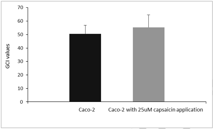
AgNOR detection The stained slides were viewed via a light microscope (Nikon Labophot 2, Olympus model CHK), and cultured Caco-2 cell line were photographed using a digital camera (ZEISS AxioCam ICc5). Captured images were transferred to image processing software (ImageJ version 1.47t, National Institutes of Health, Bethesda, Maryland, USA), and the staining intensity of AgNOR proteins were performed. One hundred nuclei per group were evaluated. The staining intensity of AgNOR proteins by silver in interphase nuclei of Caco-2 cell line calculated and expressed as gray colour index (GCI) value. The mean GCI values of staining intensity by silver of AgNOR protein in interphase nuclei of Caco-2 cell line with or without capsaicin application have been shown in the Table 1. It was shown that Caco-2 cell line are darker stained by silver and its staining intensity have lower GCI value. Conversly, the Caco-2 cell line with 25uM capsaicin application are stained lightly by silver and its staining intensity has higher GCI value (Figure 1).
Statistical analysis
| ||||||
|
RESULTS
| ||||||
|
The GCI values of AgNOR proteins are 50.594±6.381 and 55.403±9.178 for cultured Caco-2 cell line and cultured Caco-2 cell line with 25 uM capsaicin application, respectively. When groups were compared one-by-one using Mann–Whitney U Test, the GCI value of cultured Caco-2 cell line was significantly lower than cultured Caco-2 cell line with 25uM capsaicin application (Z=-4.293; p=0.000) (Table 1) (Figure 1). | ||||||
| ||||||
| ||||||
|
DISCUSSION
| ||||||
|
Although there are important advances for the management and treatment of the disease, the cancer depending deaths have been increasing, too. So different treatment strategies such as phytothrerapy have been used. Natural therapeutic agents obtained from different medical and aromatic plants such as capsaicin are used for the treatment of the disease. It is discussed that Capsaisin is a cancer predisposing factor besides its protective effect against cancer and it is important factors in the usage of dose, duration and cell type in the direction of these two effects [27], [28], [29]. In addition to anticarcinogenic effect, the capsaicin has antiangiogenic activity, antimutagenic and antigenotoxic effects [30]. Capsaicin has an anti-proliferative effect on xenograft mouse models and cancer cell lines by stopping the cell cycle in G0/G1 phases and especially inducing tumor cell apoptosis. Therefore it is an important class of therapeutics agents [31], [32]. To our knowledge, there is no research in the literature on the comparison of AgNOR protein staining intensity by using silver staining technique between cultured human Caco-2 cell line and cultured human Caco-2 cell line with 25 uM capsaicin application. Because of the AgNOR proteins amount increased in metabolically active cells, we intend to comparison of staining intensity of AgNOR proteins amount in Caco-2 cell line with and without capsaicin application. As described in material-methods section, the staining intensity by silver of AgNOR proteins has inverse correlation with the GCI value. So, Caco-2 cells stained as darker with silver have more AgNOR protein content but have lower GCI value. Also, Caco-2 cells stained as lighter with silver have less AgNOR protein content but have higher GCI value (Figure 1). The staining intensity of AgNOR proteins by silver show the rRNA transcription activity of the cell [33]. Thus, the knowledge about the activation or repression of rRNA genes can be obtained using AgNOR staining technique. Due to the AgNOR staining intensity can be used as a marker for ribosome biogenesis amount, comparison of this value between Caco-2 cell line and Caco-2 cell line with capsaicin application may give information about the effect of capsaicin on the rRNA genes regulation and ribosome biogenesis. Our current study show that staining intensity of AgNOR protein in Caco-2 cell line is significantly higher (lower GCI value) than the Caco-2 cell line with capsaicin application (higher GCI value) (p=0.001). Because the AgNOR proteins indicate proliferation ratio of the cells, it can be said that the capsaicin has potantially cancer protective effects. Also AgNOR detection method is cheap, simplicity and cost-effectiveness. In metabolically active cells, in addition to cellular morphology, gene expression and its yields are altered, too. Thus, in addition to detection of AgNOR size, number, shape and scatter; the detection of the staining intensity of this proteins are important to obtain better and more confirmable knowledge about the behavioral, metabolical activity and protein synthesis capacity of the cells [34]. Thus it can be said that AgNOR staining technique may be used to obtain knowledge about the anticancer effect of the phytotherapeutic agents and its most reliable dose for therapeutic uses. The staining intensity of AgNOR proteins helps determine the proliferation rate of the cells. An increase in the staining intensity of AgNOR proteins is associated with an increase in cellular activity. In our study, it was shown that the AgNOR proteins amount, used as a marker for proliferation rate, decrease depending on the capsaicin treatment. As a result, the current study showed capsaicin has potentially phytotherapeutic agents for cultured human colon adenocarcinoma cell line with 25uM Capsaicin application. So AgNOR staining intensity may be used as indirect indicators to obtain knowledge about the anticancer effects of the different phytotherapeutic agents such as capsaicin. New studies including different doses of the phytotherapeutic agents such as capsaicin are also needed to have more certain knowledge about the current topic. | ||||||
|
CONCLUSION
| ||||||
|
The current study showed capsaicin has potentially phytotherapeutic agents for cultured human colon adenocarcinoma cell line with 25 uM Capsaicin application. So AgNOR staining intensity may be used as indirect indicators to obtain knowledge about the anticancer effects of the different phytotherapeutic agents such as capsaicin. New studies including different doses of the phytotherapeutic agents such as capsaicin are also needed to have more certain knowledge about the current topic. | ||||||
|
REFERENCES
| ||||||
| ||||||


|
[HTML Abstract]
[PDF Full Text]
|
|
Author Contributions
Recep Eröz – Substantial contributions to conception and design, Acquisition of data, Analysis and interpretation of data, Drafting the article, Revising it critically for important intellectual content, Final approval of the version to be published Merve Aipay – Substantial contributions to conception and design, Acquisition of data, Analysis and interpretation of data, Drafting the article, Revising it critically for important intellectual content, Final approval of the version to be published |
|
Guarantor of Submission
The corresponding author is the guarantor of submission. |
|
Source of Support
None |
|
Consent Statement
Written informed consent was obtained from the patient for publication of this study. |
|
Conflict of Interest
Author declares no conflict of interest. |
|
Copyright
© 2018 Recep Eröz et al. This article is distributed under the terms of Creative Commons Attribution License which permits unrestricted use, distribution and reproduction in any medium provided the original author(s) and original publisher are properly credited. Please see the copyright policy on the journal website for more information. |
|
|