| Table of Contents |  |
|
Original Article
| ||||||
| Angiogenic cytokines: IL-6, sIL-6R, TNF-α, sVCAM-1, and PDGF-AB in multiple myeloma patients depending on the stage of the disease | ||||||
| Joanna Kamińska1, Olga M. Koper1, Violetta Dymicka-Piekarska2, Elżbieta Motybel3, Janusz Kłoczko4, Halina Kemona5 | ||||||
|
1PhD, Researcher, Department of Clinical Laboratory Diagnostics, Medical University of Bialystok, Poland.
2Assistant Professor, Researcher, Department of Clinical Laboratory Diagnostics, Medical University of Bialystok, Poland. 3MS, Researcher, Department of Clinical Laboratory Diagnostics, Medical University of Bialystok, Poland. 4Head of the Department, Department of Hematology, Clinical Hospital of the Medical University of Bialystok, Poland. 5Head of the Department, Department of Clinical Laboratory Diagnostics, Medical University of Bialystok, Poland. | ||||||
| ||||||
|
[HTML Abstract]
[PDF Full Text]
[Print This Article]
[Similar article in Pumed] [Similar article in Google Scholar] |


| How to cite this article |
| Kamińska J, Koper OM, Dymicka-Piekarska V, Motybel E, Kloczko J, Kemona H. Angiogenic cytokines: IL-6, sIL-6R, TNF-α, sVCAM-1, and PDGF-AB in multiple myeloma patients depending on the stage of the disease. Edorium J Tumor Bio 2015;2:11–19. |
|
Abstract
|
|
Aims:
Angiogenesis plays an important role for growth, progression and metastasis of various cancers, including multiple myeloma (MM). Therefore, the aim of the study was the evaluation of concentrations of chosen angiogenic cytokines: interleukin 6 (IL-6), its soluble receptor (sIL-6R), tumor necrosis factor-α (TNF-α), soluble vascular cell adhesion molecule-1 (sVCAM-1), and platelet-derived growth factor-AB (PDGF-AB) in patient with newly diagnosed MM depending on the stage of the disease and as compared to the control group.
Methods: The study group consisted of newly diagnosed MM patients prior to treatment and categorized depending on the Durie and Salmon staging system. The controls consisted of healthy subjects. Angiogenic cytokines were determined with the use of ELISA method. Results: Serum concentrations of all angiogenic cytokines analyzed were significantly higher in the whole study group as compared to the controls. Moreover, concentrations of all proteins tested significantly increasing with the stage of MM. Additionally, all cytokines tested positively correlated with the percentage of plasma cells in the bone marrow. The areas under the ROC curves (AUCs) for all cytokines analyzed were significantly higher than AUC=0.500. Conclusion: Concentrations of angiogenic cytokines analyzed were significantly higher in MM patients as compared to the healthy subjects. Additionally, the concentrations of proteins tested were significantly increasing with the stage of the disease. Since the progression of MM proceeds at the same time as angiogenesis it allowed us to hypothesize that cytokines analyzed take part in bone marrow neovascularization. The areas under ROC curves analysis may indicate that these cytokines have potential clinical significance in MM. | |
|
Keywords:
Angiogenic cytokines, Multiple myeloma, Platelet-derived growth factor-AB, Soluble vascular cell adhesion molecule-1
| |
|
Introduction
| ||||||
|
Multiple myeloma (MM) accounts 1–2% of all human cancers and approximately 10 percent of all hematological malignancies. MM is associated with clonal proliferation of plasma cells in the bone marrow, the presence of monoclonal protein (a single abnormal immunoglobulin) in the serum and/or in urine, and osteolytic bone lesion [1] [2]. Angiogenesis is a multistep process of the formation of new blood vessels during embryonic growth, tissue healing, and in the female in regeneration of endometrium during menstrual cycle [3]. Angiogenenesis also occurs in growth and metastases of solid tumors. It is also important in hematological malignancies, such as MM [4] [5]. The measuring of microvessel density (MVD) is an objective method used to the evaluation of neovascularization in bone marrow [5]. In MM, angiogenesis was defined as a prognostic factor [6]. Vacca et al. revealed a high correlation between the increased bone marrow angiogenesis, estimated by measuring of MVD, and the proliferating fraction of plasma cells (estimated as labeling index) in Monoclonal Gammopathy of Undetermined Significance (MGUS) and MM patients. On the basis of obtained results authors suggested that MM is angiogenesis-dependent [4] . It is well established that various pro-inflammatory cytokines and growth factors secreted by bone marrow microenvironment cells and malignant plasma cells play an important role in the multistep process of angiogenesis [7] [8] [9]. These proteins include interleukins (e.g.: IL-1, -6, -8, -10) and mitogenic growth factors (e.g.: vascular endothelial growth factor, VEGF; basic fibroblast growth factor, b-FGF; tumor necrosis factor-alpha, TNF-α; insulin-like growth factor 1, IGF-1; platelet derived growth factor-AB, PDGF-AB; stem cell factor, SCF) and have direct impact on growth, survival and metastasis of plasma cells [10] [11]. Studies showed positive correlations between serum concentrations of chosen pro-angiogenic cytokines and MVD [12] . Therefore, the aim of the current study was to assess the serum concentrations of chosen angiogenic cytokines, such as: interleukin 6 (IL-6) and its soluble receptor (sIL-6R), tumor necrosis factor-α (TNFα), soluble vascular cell adhesion molecule-1 (sVCAM-1), and platelet-derived growth factor-AB (PDGF-AB) in patients with newly diagnosed MM as compared to the healthy controls. Moreover serum concentrations of above-mentioned cytokines were analyzed depending on the stage of the disease and other markers of the MM activity: β2-microglobulin (β2M), albumin (Alb) concentrations, lactate dehydrogenase activity (LDH), and the percentage of plasma cells in the bone marrow (% of plasma cells). Additionally, the areas under receiver operating characteristic (ROC) curves (AUCs) for all proteins tested were assessed. | ||||||
|
Materials and Methods
| ||||||
|
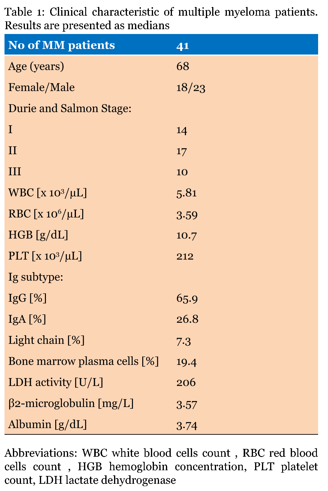
The study group (MM) included 41 patients (18 females and 23 males, mean age 68 years, range 47–86 years) with newly diagnosed MM, prior to treatment. Patients were diagnosed at the Department of Hematology of the Clinical Hospital of the Medical University of Bialystok according to the World Health Organization (WHO) criteria, including: an increased number of abnormal, atypical or immature plasma cells in the bone marrow or histological proof of plasmocytoma; the presence of an M protein in the serum and/or in urine; bone lesions [13]. The patients were categorized depending on the stage of the disease according to the Durie and Salmon staging system [14]: I stage, II stage, and III stage. Table 1 presents the clinical characteristic of the study group. The control group (C) consisted of 30 healthy volunteers (15 F/15 M, mean age 66 years, range 45–77 years). The study was approved by the Bioethics Committee on human research of the Medical University of Bialystok (permission number: R-I-002/112/2009). All the patients gave they written informed consent to participate in the study. Blood samples from the patients group and the controls were drawn between 6–7 o'clock in the morning following a fasting period of 10–12 hours. Tubes with the blood collected without anticoagulant were allowed to clot for 30 minutes before centrifugation for 15 minutes at 1000 xg, obtained sera were stored at –750C until further analysis. The IL-6, sIL-6R, TNF-α, sVCAM-1, and PDGF-AB concentrations in the sera were measured with the use of commercially available ELISAs (Quantikine ELISA ® R&D Systems Inc., Abingdon, United Kingdom) according to the manufacturer's instructions. The β2-microglobulin and albumin concentrations were measured with the use of immunonephelometry method on the BN* II (Siemens, Berlin, Germany). The serum lactate dehydrogenase activity (LDH) was determined spectrophotometrically on the ARCHITECT c Systems™ (ABBOTT Park, IL, USA). The percentage of plasma cells in the bone marrow was evaluated in bone marrow smears under light microscopy. Statistical analysis | ||||||
| ||||||
|
Results | ||||||
|
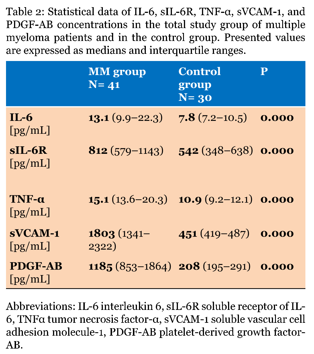
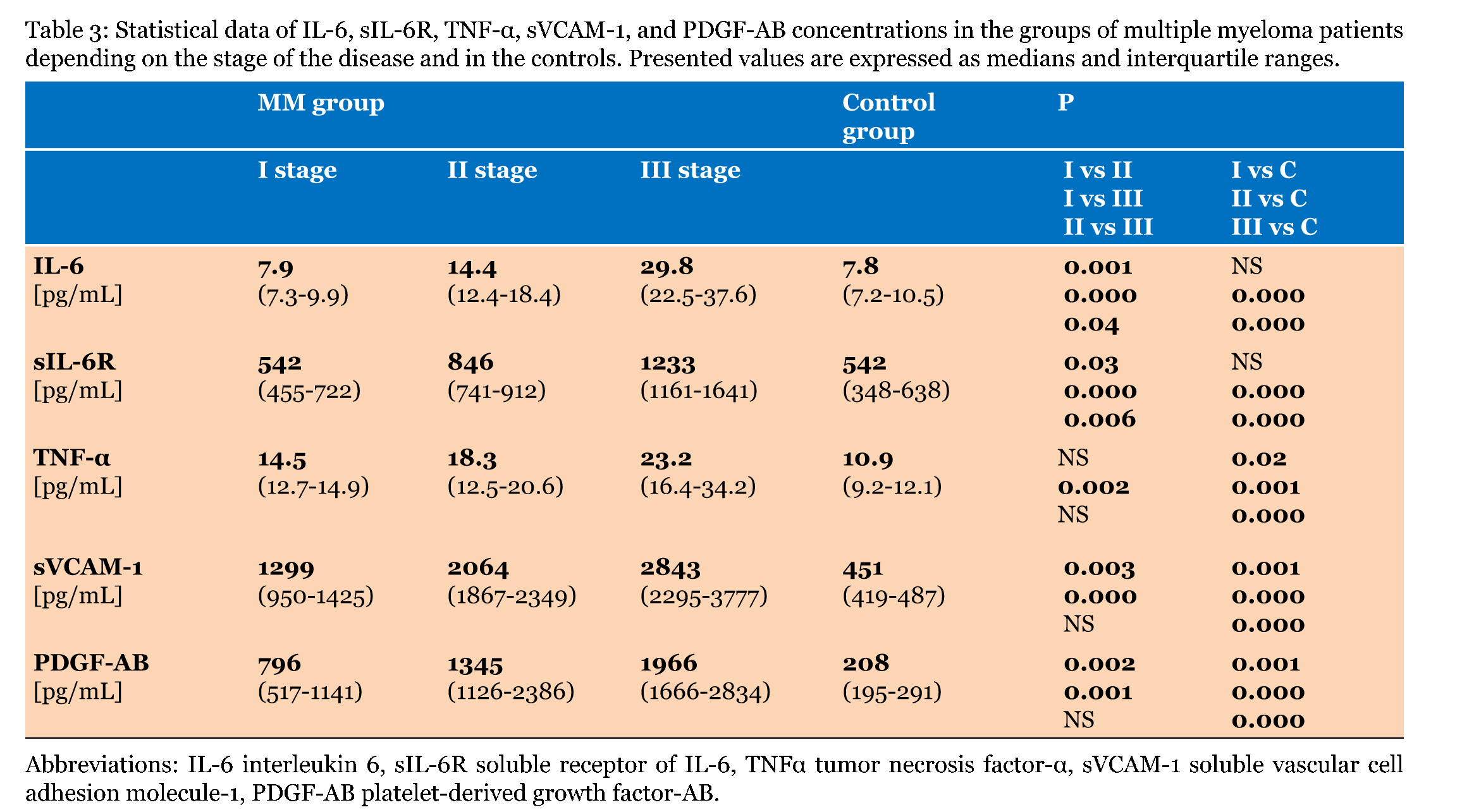
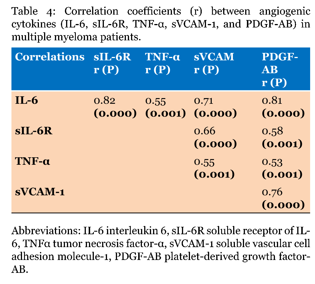
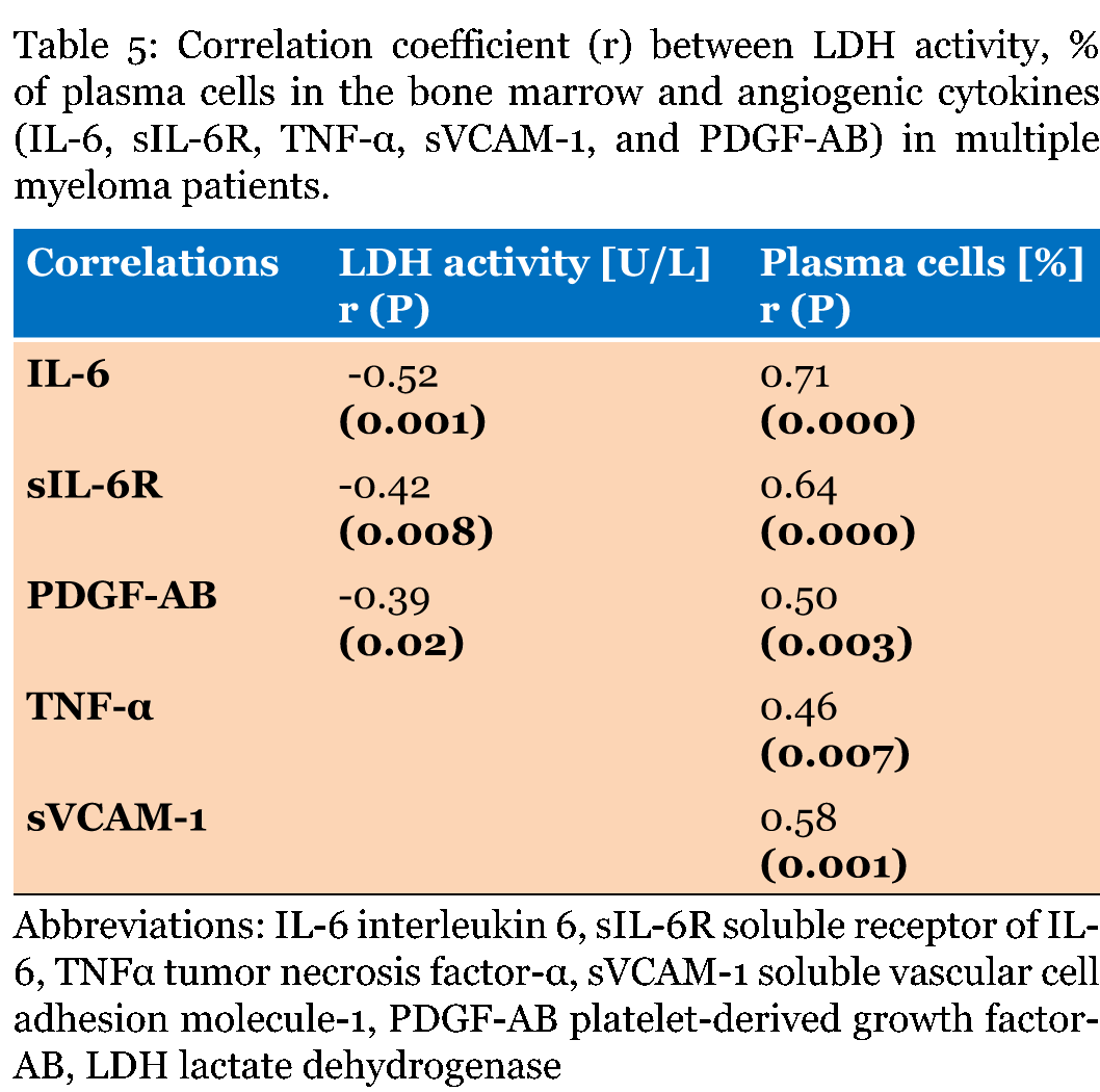
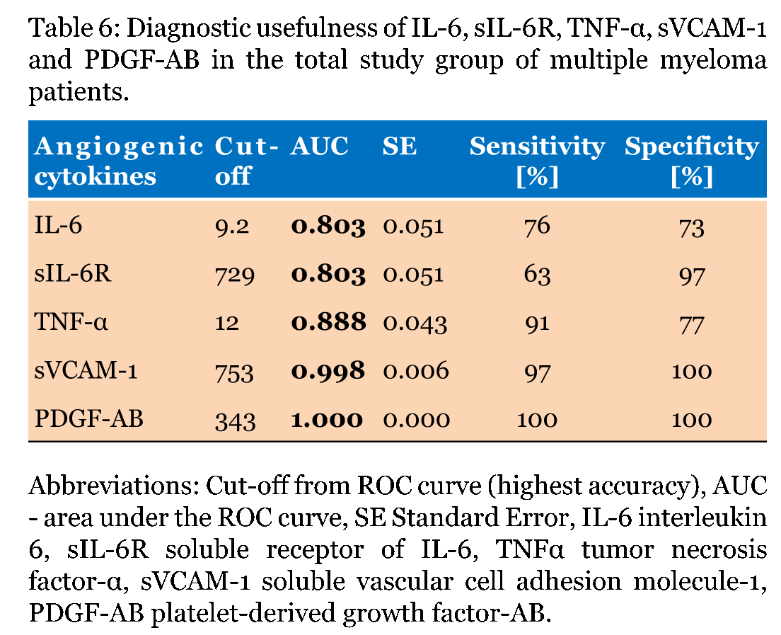
Serum concentrations of all angiogenic cytokines tested (IL-6, sIL-6R, TNF-α, sVCAM-1, and PDGF-AB) in the whole study group were significantly higher as compared to the control group. Median PDGF-AB concentration was approximately 6-fold higher in the MM group as compared to the healthy subjects (Table 2). The analysis of median concentrations depending on the stage of the disease revealed that the highest medians were in the III stage of MM in case of all proteins tested. The post-hoc tests revealed that the statistically significant differences were not observed only between II versus III stage in case of TNF-α, sVCAM-1, and PDGF-AB and between I versus II stage in case of TNF-α (Table 3). Table 4 presents the correlation coefficients between IL-6, sIL-6R, TNF-α, sVCAM-1, and PDGF-AB in MM patients. IL-6 and sVCAM-1 were significantly positively correlated with sIL-6R, TNF-α, sVCAM-1, and PDGF-AB. sIL-6R as well as TNF-α revealed a correlation coefficient with IL-6, sVCAM-1, and PDGF-AB (Table 4). The Spearman's rank method did not reveal correlations between angiogenic cytokines tested and β2-microglobulin as well as albumin concentrations (data not shown). The percentage of plasma cells in the bone marrow positively correlated with all cytokines analyzed, whereas the LDH activity negatively correlated with IL-6, sIL-6R, and PDGF-AB concentrations (Table 5). Table 6 summarizes the results of diagnostic usefulness of angiogenic cytokines tested. AUCs for all proteins tested were significantly higher than AUC=0.500. Interestingly the highest sensitivity and specificity showed PDGF-AB (100%); also sVCAM-1 revealed high sensitivity and specificity (97% and 100%, respectively). Moreover the biggest AUCs were observed for PDGF-AB and sVCAM-1. The AUCs for IL-6 and sIL-6R were identical (Table 6). | ||||||
|
| ||||||
|
| ||||||
| ||||||
| ||||||
|
| ||||||
| ||||||
|
Discussion
| ||||||
|
Current study revealed that serum concentrations of IL-6, sIL-6R, TNF-α, sVCAM-1, and PDGF-AB, assessed using ELISA method, were significantly higher in MM patients as compared to the healthy subjects. Additionally, the concentrations of angiogenic cytokines were significantly increasing with the stage of the disease, which is in agreement with findings of other authors [9] [15]. IL-6 is produced by myeloma cells by both autocrine and paracrine mechanisms [16]. In MM, IL-6 takes part not only in the pathogenesis of the disease but also it is involved in the activation of angiogenic pathways [17]. The secretion of angiogenic growth factors by plasma cells is stimulated by IL-6 [15] [18] . IL-6 mediates its effect through a cell surface receptor build of the IL-6Rβ (CD 130) and the specific ligand-binding protein (IL-6Ra). IL-6Ra exists in membrane-bound form (CD126) and in soluble form (sIL-6Ra). IL-6 can bind to IL-6Ra to generate complex of IL-6/IL-6Ra/CD130, leading to the activation of the intracellular signaling cascade. Therefore, cells that express only CD130 are sensitive only to exogenous IL-6/IL-6Ra chains complex [18] [19]. CD130 is presented on the surface of most of the cells, while the cell surface receptor for IL-6 is expressed only on the membranes of a single cells, e.g.: megakaryocytes, hepatocytes, neutrophils, monocyte/macrophages, and lymphocytes [20]. In the present study, strong positive correlation coefficient between IL-6 and sIL-6R was revealed. Moreover, the concentrations of sIL-6R were 1.5-fold higher in MM patients as compared to the healthy individuals and significantly higher in the III stage of the disease as compared to the I and II stage of MM, which may indicate that both IL-6 and its soluble receptor might be recognized as a markers of poor prognosis. Our results are in line with findings of Pulkki et al., which indicated that sIL-6R was a poor prognostic factor in MM [21]. Furthermore, in the current study LDH activity significantly correlated with IL-6 and sIL-6R. It should be emphasized that increased LDH activity is a biochemical predictor of poor prognosis in MM [22]. We also revealed that the IL-6 and sIL-6R significantly correlated with the percentage of the plasma cells in the bone marrow. TNF-α is also involved in MM angiogenesis and recognized as an important factor in a survival for human myeloma cells. In MM, TNF-α is synthesized by stromal and plasma cells [23]. Moreover, it triggers the secretion of proangiogenic cytokines, including IL-6, VEGF [15] [24]. TNF-α and IL-6 stimulate migration of endothelial cells. Synergistic common action of both mentioned cytokines cause significantly higher migration of endothelial cells than the action of each cytokine separately [10] . TNF-α also enhance the transendothelial migration of myeloma cells [25]. In the available literature only, the study of Hatjiharissi et al. concerns the evaluation of TNF-α in MM patients as compared to healthy subjects, in which no significant difference was found [26]. The result is in disagreement with our findings. The discrepancy between these two study could be explained by different number of subjects included into the patients group (in our study MM group included 41 patients; in the study of Hatjiharissi et al. 25 patients). In the current study, the positive correlation between IL-6 and TNF-α was found, which is with agreement with the investigation of Zdzisinska et al. [27]. It allows us to hypothesize that increased secretion of IL-6 and TNF-α may influence angiogenesis because the progression of MM proceeds at the same time as angiogenesis. Therefore, the inhibition of neovascularyzation by effective chemotherapy would be especially important in the reduction of MM growth [15]. Vascular cell adhesion molecule 1 (VCAM-1) is a transmembrane glycoprotein expressed on vascular endothelial and tumor cells in response to inflammatory cytokines. In the circulation, VCAM-1 is presented in soluble form (sVCAM-1) [28]. Increased sVCAM-1 concentrations were found in various malignancies, such as breast cancer and gastric cancer [28] [29]. It is suggested that leukocyte-endothelial adhesion and neoangiogenesis are linked, because VCAM-1 expression is induced by TNF-α and VEGF, which are angiogenic cytokines [28] [30]. To the best of our knowledge the evaluation of concentrations of adhesion molecules in MM were investigated only by Scudla et al. [31]. The results of Scudla et al. are in agreement with our study, in which significantly higher sVCAM-1 concentrations in MM patients as compared to the control group was observed; sVCAM-1 concentrations were also significantly increasing with the stage of the disease. Additionally, the correlations coefficient between sVCAM-1 and all angiogenic cytokines tested were observed. Obtained results allow us to hypothesize that the increased sVCAM-1 concentrations, beside the pro-angiogenic cytokines and growth factors concentrations, may be used as a marker of angiogenesis in MM. Growth factors also have an angiogenic potential. The platelet-derived growth factor (PDGF) family consists of four different molecules: PDGF-A,-B,-C, and -D, which may form either homodimers (AA, BB, CC, DD) or heterodimers (AB) [32] [33] . PDGF is expressed by various cells (e.g., epithelial and endothelial cells, macrophages, nervous tissue, vascular and smooth muscle cells, and bone marrow stromal cells), and released by activated platelets and megakaryocytes [34] [35] . PDGF are a major mitogens for cancer development by autocrine and paracrine signaling binding to the PDGFR-a and -β receptors [36]. PDGF-AB is considered as a potent stimulator of angiogenesis in many solid tumours and haematological malignancies, including MM [9]. PDGF-AB influences tumor angiogenesis by direct induction of VEGF production [37]. In the current study, serum PDGF-AB concentrations in MM patients were significantly higher as compared to the healthy subjects and were increasing with the stage of the disease. Furthermore, positive correlations between PDGF-AB and all cytokines tested, as well as the LDH activity and the percentage of plasma cells in the bone marrow were observed. Our study is in the line with the reports of Tsirakis et al., which also revealed positive correlation between serum PDGF-AB and IL-6 concentrations [9]. Our findings, supported by the results of Tsirakis et al., support the hypothesis that increased PDGF-AB concentrations play an important role in angiogenesis in MM [9]. It should be emphasized that this is the first study estimating the diagnostic usefulness of IL-6, sIL-6R, TNF-α, sVCAM-1, PDGF-AB in MM patients. The AUCs analysis revealed that areas under ROC curves for all angiogenic proteins tested were significantly higher than AUC=0.500, which may indicate that these cytokines have potential clinical significance in MM. Among all cytokines tested the largest area under ROC curve and thereby the greatest potential clinical usefulness in MM had sVCAM- 1 and PDGF-AB. | ||||||
|
Conclusion
| ||||||
|
In conclusion, increased IL-6, sIL-6R, TNF-α, sVCAM-1, and PDGF-AB concentrations in MM patients as compared to the healthy subjects may support the hypothesis that these proteins play an important role MM. Additionally, the serum concentrations of all proteins tested were significantly increasing with the stage of the disease, which may indicate that above-mentioned cytokines take part in the progression of MM. It allows us to hypothesize that cytokines analyzed take part in bone marrow neovascularization because the progression of MM proceeds at the same time as angiogenesis. The largest areas under ROC curves were for sVCAM- 1 and PDGF-AB, which may indicate that these proteins have the highest potential clinical usefulness in MM. Certainly, further studies, on larger study group, are needed to explain whether IL-6, sIL-6R, TNF-α, sVCAM-1, and PDGF-AB may be utilized as potential tools for the evaluation of neoangiogenesis in MM patients. | ||||||
|
References
| ||||||
| ||||||


|
[HTML Abstract]
[PDF Full Text]
|
|
Author Contributions:
Joanna Kamińska – Substantial contributions to conception and design, Acquisition of data, Analysis and interpretation of data, Drafting the article, Revising it critically for important intellectual content, Final approval of the version to be published Olga M. Koper – Substantial contributions to conception and design, Analysis and interpretation of data, Drafting the article, Revising it critically for important intellectual content, Final approval of the version to be published Violetta Dymicka-Piekarska – Analysis and interpretation of data, Revising it critically for important intellectual content, Final approval of the version to be published Elżbieta Motybel – Analysis and interpretation of data, Revising it critically for important intellectual content, Final approval of the version to be published Janusz Kłoczko – Analysis and interpretation of data, Revising it critically for important intellectual content, Final approval of the version to be published Halina Kemona – Analysis and interpretation of data, Revising it critically for important intellectual content, Final approval of the version to be published |
|
Guarantor of submission
The corresponding author is the guarantor of submission. |
|
Source of support
None |
|
Conflict of interest
Authors declare no conflict of interest. |
|
Copyright
© 2015 Joanna Kamińska et al. This article is distributed under the terms of Creative Commons Attribution License which permits unrestricted use, distribution and reproduction in any medium provided the original author(s) and original publisher are properly credited. Please see the copyright policy on the journal website for more information. |
|
|
|
About The Authors
| |||
| |||
| |||
| |||
| |||
| |||
| |||