| Table of Contents |  |
|
Original Article
| ||||||
| Clinicopathological features and prognostic implications of loss of K5 and gain of K1, K8 and K18 in oral potentially malignant lesions and squamous cell carcinomas: An immunohistochemical analysis | ||||||
| Sawant Sharada1, Vaidya Milind2, Chaukar Devendra3, Gangadaran Prakash4, Singh Archana5, Rajadhyax Siddheshwar6, Kannan Sadhana7, Padmavathi A8, Kane Shubhada9, Pagare Sandeep10, Kannan Ranganathan11, D'Cruz Anil12 | ||||||
|
1MSc, Scientific Officer, Vaidya Lab., Advanced Centre for Treatment, Research and Education in Cancer (ACTREC), Tata Memorial Centre, Kharghar, Navi Mumbai, 410 210, Maharashtra, India.
2PhD, Scientific Officer, Vaidya Lab., Advanced Centre for Treatment, Research and Education in Cancer (ACTREC), Tata Memorial Centre, Kharghar, Navi Mumbai, 410 210, Maharashtra, India. 3MS, Professor and Surgeon, Oral Surgery, Head and Neck Unit, Tata Memorial Hospital, Parel, Mumbai, 400 012, Maharashtra, India. 4MSc, Post graduate student, Department of Nuclear Medicine, Kyungpook National University, Daegu, Republic of Korea, 700-721. 5MSc, Research Assistant, Advanced Centre for Treatment, Research and Education in Cancer (ACTREC), Tata Memorial Centre, Kharghar, Navi Mumbai, 410 210, Maharashtra, India. 6MSc, Research Assistant, Advanced Centre for Treatment, Research and Education in Cancer (ACTREC), Tata Memorial Centre, Kharghar, Navi Mumbai, 410 210, Maharashtra, India. 7MSc, Program Manager, Epidemiology and Clinical Trials Unit, Clinical Research Centre, Advanced Centre for Treatment, Research and Education in Cancer (ACTREC), Kharghar, Navi Mumbai, Maharashtra, India. 8MSc, ad-hoc statistician, Epidemiology and Clinical Trials Unit, Clinical Research Centre, Advanced Centre for Treatment, Research and Education in Cancer (ACTREC), Kharghar, Navi Mumbai, Maharashtra, India. 9MD, Professor and Pathologist, Pathology Department, Tata Memorial Hospital, Parel, Mumbai, 400 012, Maharashtra, India. 10MDS, Professor and Chief, Department of Oral Medicine & Radiology, D.Y. Patil Dental College and Hospital, Sector 7, Nerul, Navi-Mumbai-400706, Maharashtra, India. 11MDS, Professor and Chief, Department of Oral and Maxillofacial Pathology, Ragas Dental College and Hospital, Uthandi, 600096 Chennai, India. 12MS, Professor and Chief, Oral Surgery, Head and Neck Unit, Tata Memorial Hospital, Parel, Mumbai, 400 012, Maharashtra, India. | ||||||
| ||||||
|
[HTML Abstract]
[PDF Full Text]
[Print This Article]
[Similar article in Pumed] [Similar article in Google Scholar] |


| How to cite this article |
| Sharada S, Milind V, Devendra C, Prakash G, Archana S, Siddheshwar R, Sadhana K, Padmavathi A, Shubhada K, Sandeep P, Ranganathan K, Anil DC. Clinicopathological features and prognostic implications of loss of K5 and gain of K1, K8 and K18 in oral potentially malignant lesions and squamous cell carcinomas: An immunohistochemical analysis. Edorium J Tumor Bio 2014;1:1–22. |
|
Abstract
|
|
Aims:
To analyze alterations in the expression and localization patterns of keratins-K1, K5, K8 and K18 using immunohistochemistry and correlate with clinicopathological parameters of patients with oral potentially malignant lesions and squamous cell carcinomas to evaluate diagnostic and prognostic implications of loss and gain of keratins.
Methods: Altered keratin expression pattern was investigated using immunohistochemistry in tissues of oral normal mucosa (n=10), leukoplakia (n=50), submucous fibrosis (n=67) and tumor respective cut-margins (n=304). The prognostic significance was determined by correlating the values of these two events singly as well as in different permutations and combinations with clinicopathological parameters using univariate and multivariate analyzes. Results: Loss of K5 and aberrant expression of K1, K8 and K18 were seen in oral premalignant lesions as well as tumor tissues in comparison to normal oral mucosal tissues. Non-expression of K5 (p=0.003), and aberrant expression of K1 (p<0.001), K8 (p=0.001), and K18 (p=0.004), independently significantly correlated with clinicopathological progressive grade of oral premalignant disorders as well as some of the clinicopathological factors of patients with oral cancer. The univariate and multivariate analysis showed the significance of combination of keratin markers (K1, K8, K18) on overall survival and local recurrence free survival of patients with oral cancer. The number of markers combined together has increased the risk of recurrence significantly (p<0.0001). Conclusion: These findings suggest, loss and gain of keratins could serve as surrogate markers for the diagnosis of oral potentially malignant disorders and may also have prognostic value in patients with oral cancer. | |
|
Keywords:
Keratin biomarkers, Leukoplakia, Oral squamous cell carcinomas, Submucous fibrosis
| |
|
Introduction
| ||||||
|
Oral squamous cell carcinoma (OSCC) is the sixth largest group of malignancies worldwide [1] and the single largest malignancy in males in the Indian subcontinent [2]. In Indian scenario, major contributory factors for the high incidence of oral cancer are the habits of chewing tobacco, areca nut, and other allied products coupled with alcohol consumption, lower socioeconomic status, and poor oral hygiene [3]. It is well accepted fact that in India, most invasive oral cancers arise from potentially malignant disorders of oral mucosa such as leukoplakia, erythroplakia and submucous fibrosis (SMF). The incidence of oral cancer in subjects with tobacco habits is 50-fold higher as compared to tobacco non-users [3]. The follow-up data show that the risk of malignant transformation of oral leukoplakia varies widely between 0.5% and 20% [4], whereas for SMF, it ranges between 4.5% and 7.6% [5]. Dysplasia is being currently used as standard predictive parameter to predict the risk for the conversion of potentially malignant lesions into frank malignancy [5] [6]. However, histopathological assessment is rather subjective [7] and the existing imaging modalities are also not sensitive enough to predict the risk of malignant conversion [8]. Despite the advances in surgical and therapeutic modalities, the prognosis for patients with OSCC remains poor and the survival rate is less than 50% [9]. It is known that local recurrence and regional lymph node metastasis are major contributory factors for poor survival of oral cancer patients and have proved to be major hurdle in the management of disease. More than 40% oral cancer patients die as a result of uncontrolled local recurrence [10]. Currently, treatment decisions are based on established clinicopathological parameters like the TNM classification. However, tumor progression seems to be a multifactorial and multistep process [11], where, accumulation of genetic defects is reflected into molecular alterations which further lead to the development of cancer. Since molecular changes occur before cellular or clinical changes are evident, detection of these molecular changes would ideally allow early diagnosis/prognosis of the disease [12]. A number of molecular markers have been proposed in the past for prognostication of oral cancer. However, their prognostic value is still not quite clear [10] [13]. Considering all these facts, it is necessary to develop other modalities as an adjunct to histodiagnosis for predicting the malignant potential of high-risk lesions as well as for the prognostication of patients with OSCC. Keratins (K) are epithelia predominant intermediate filament (IF) proteins which are expressed in a differentiation dependent, site specific and paired manner. The keratin pair of 5 and 14 is found mainly in the basal cell layer and is associated with the proliferative potential of these cells, while, the intermediary cell layers show expression of high molecular weight keratin pair of 4/13 or 1/10, which are regarded as markers of cellular differentiation. In contrast, low molecular weight keratin pair of 8/18 is normally express in glandular epithelia. K1, K8 and K18 are aberrantly expressed in buccal mucosa while K8 and K18 are aberrantly expressed in tongue tissues during oral carcinogenesis. A number of groups have studied keratin expression profile in human oral precancer as well as cancer and some consistent patterns of keratin expression have emerged from these studies [14] [15] [16] [17] [18] [19] [20]. Many of these alterations show a potential to be used as predictive markers for human oral cancer. In our previous studies, we have demonstrated non- expression of K5 and aberrant expression of K1, K8 and K18 in both oral mucosal premalignant lesions and SCC [14] [18][19][20]. These results clearly indicated the possibility of using these changes as predictive biomarkers for both oral precancerous lesions and SCC. It was necessary to use adequate sample size so as to statistically evaluate clinical significance of non-expression/aberrant expression of these proteins because of the limited sample size used in the previous studies. Previous study was conducted by one and two dimensional gel electrophoresis along with western blotting. It is difficult to prove non-expression of a protein using standard immunochemical techniques. Therefore, K5 non-expression was also studied using reverse transcriptase-polymerase chain reaction assay [14]. Although, immunohistochemistry (IHC) is a semi-quantitative technique, it gives the information about the alterations in the localization of protein which has more clinical value. Availability of more sensitive techniques and highly specific antibodies enable immunolabeling more specific. Hence, in this study our aim was to analyze alterations in the expression and localization patterns of K1, K5, K8 and K18 and correlate with clinicopathological parameters of patients with oral premalignant lesions and SCC to evaluate diagnostic and prognostic implications of altered keratins expression pattern. Our results show significant correlation of loss of K5 and gain of K1, K8, and K18 with clinical and histopathological grade of disease progression in hyperplasia/dysplasia samples. Combinations of the aberrantly expressed keratins-K1, K8 and K18 (any 1 positive, any 2 positive, and all positive) significantly correlated with overall survival and recurrence free survival of OSCC patients. Further, we also noticed the trend that the risk of tumor recurrence increased, with increased number of markers combined together (p < 0.001, sts test, trend, STATA 11.0). | ||||||
|
Materials and Methods
| ||||||
|
Patients and Tissue Specimens Clinical and Histopathological Information of Patients Five micrometers thick sections from 10% buffered formalin fixed and paraffin embedded tissues were stained with Hematoxylin and Eosin (H&E), and histopathological grading [3] was done by two independent pathologists. Histopathologically, it was confirmed that all tumors were squamous cell carcinomas (SCC). Subsequent to H&E staining, the remaining serial sections were used for IHC. This study was carried out in double blinded fashion. Immunohistochemistry Statistical analysis | ||||||
|
Results | ||||||
|
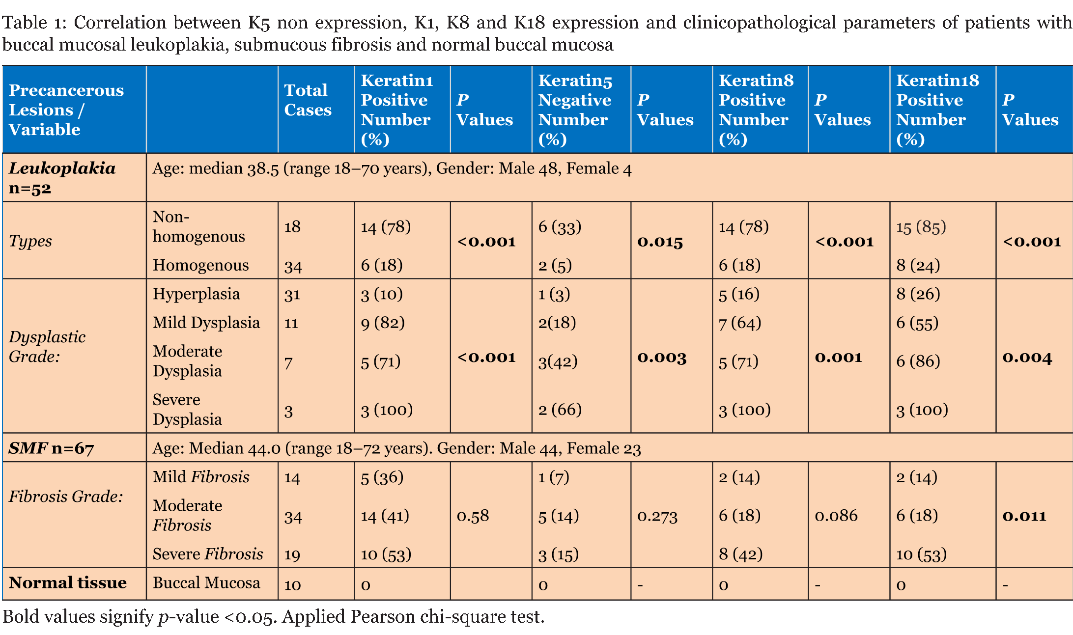
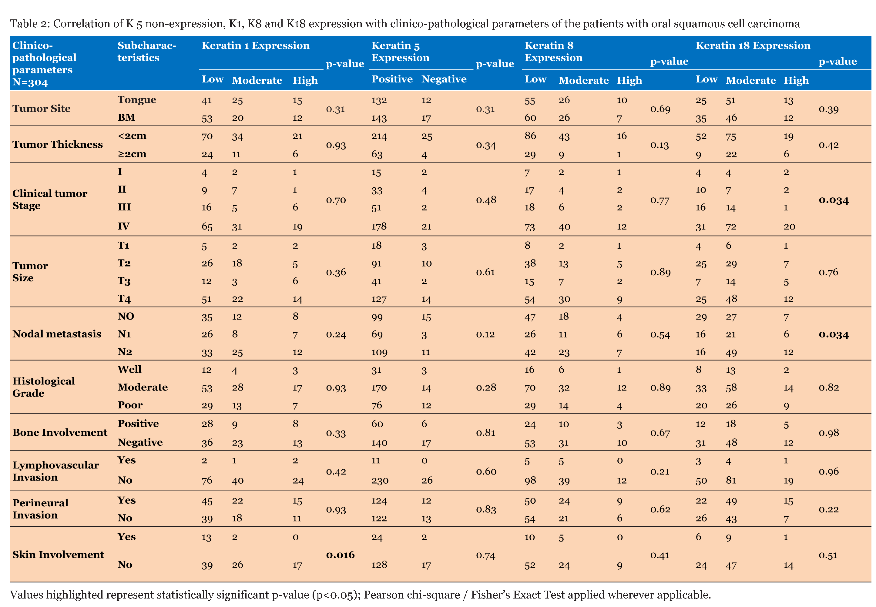
This study comprises immunohistochemical analysis of loss of K5 expression and aberrant expression of K1, K8 and K18 in the tissues with oral mucosal hyperplasia/dysplasia, fibrosis and SCC (tongue and BM) along with their respective cut margin (CM) tissues in comparison with normal oral mucosal tissues. Clinicopathological information of patients with leukoplakia and SMF and their correlation with keratin expression profile is given in (Table 1). Clinicopathological information of patients with OSCC is given in (Table 2). | ||||||
| ||||||
| ||||||
|
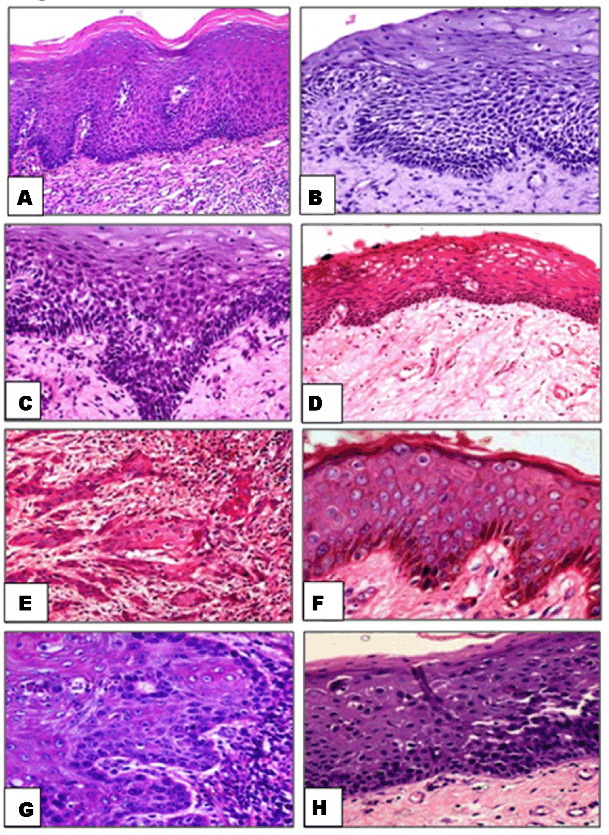
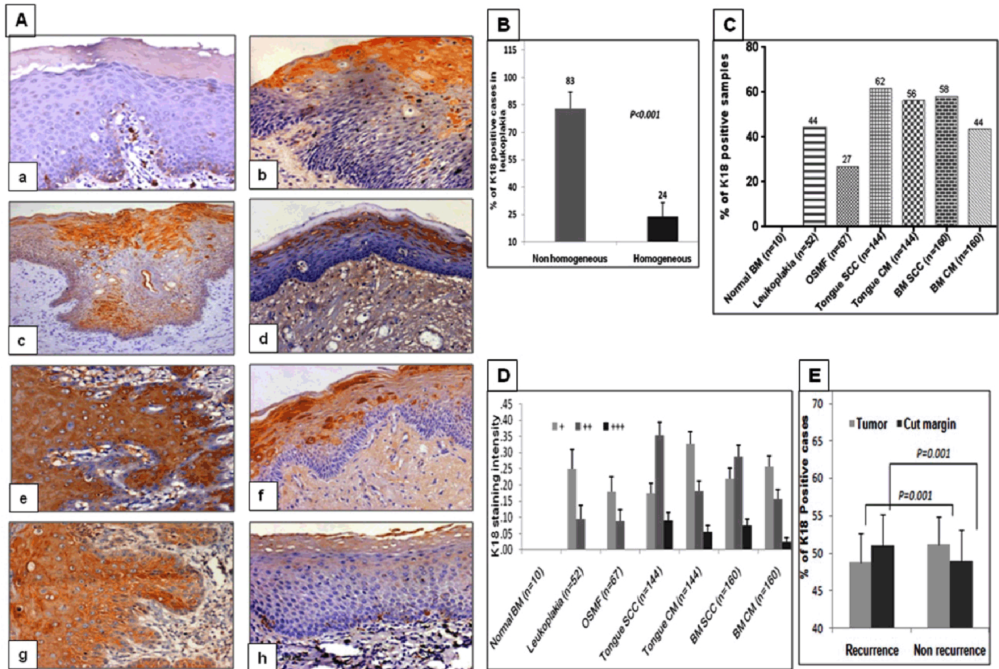
Histopathology of H&E stained sections of normal oral mucosa, mucosal hyperplasia, dysplasia, fibrosis, tongue SCC, cut margins of tongue tumors, buccal mucosal SCC and cut margins of buccal mucosal tumors is shown in (Figure 1A-H). | ||||||
|
| ||||||
|
Immunohistochemical analysis of alterations in K1, K5, K8 and K18 expression | ||||||
|
| ||||||
|
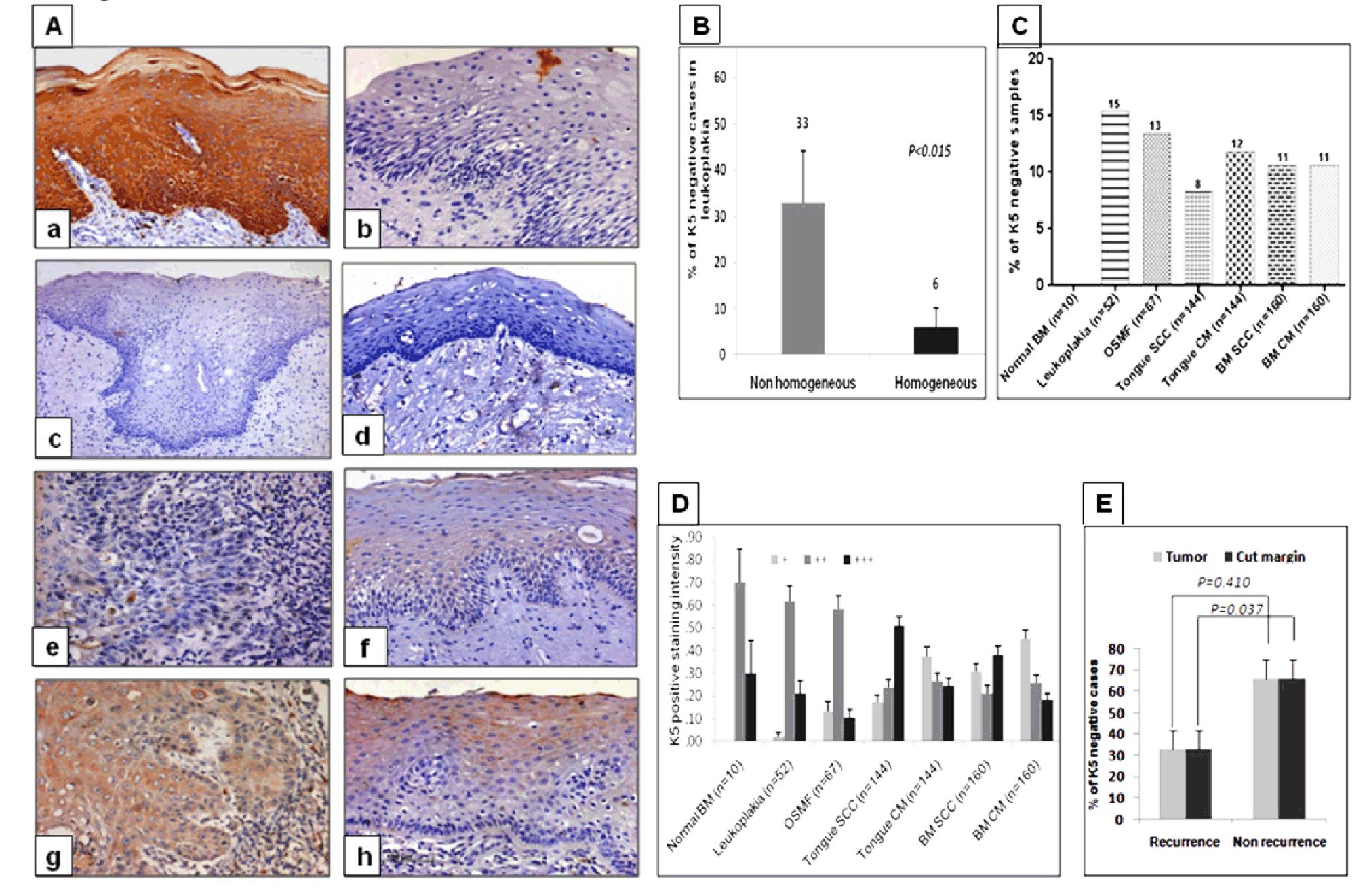
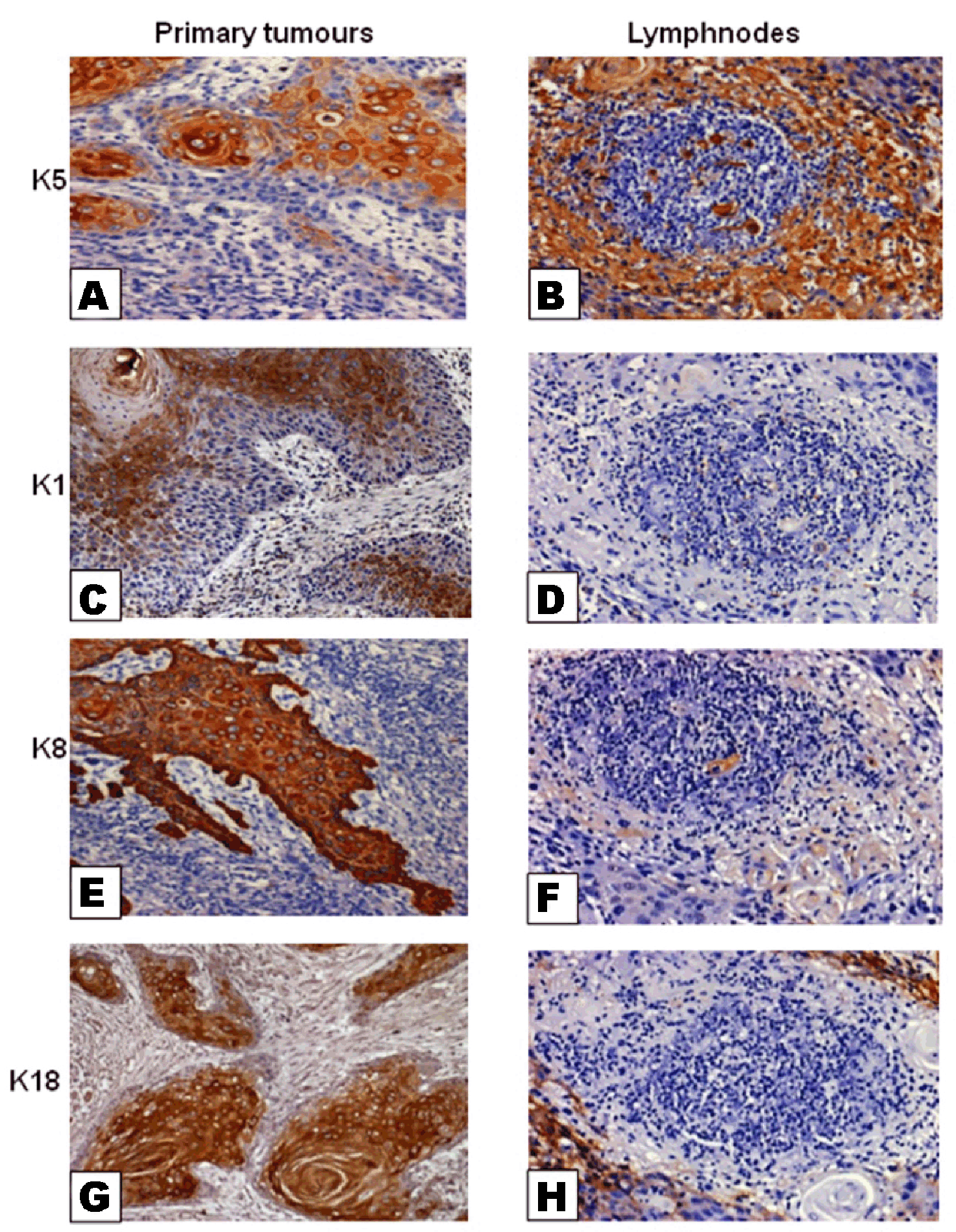
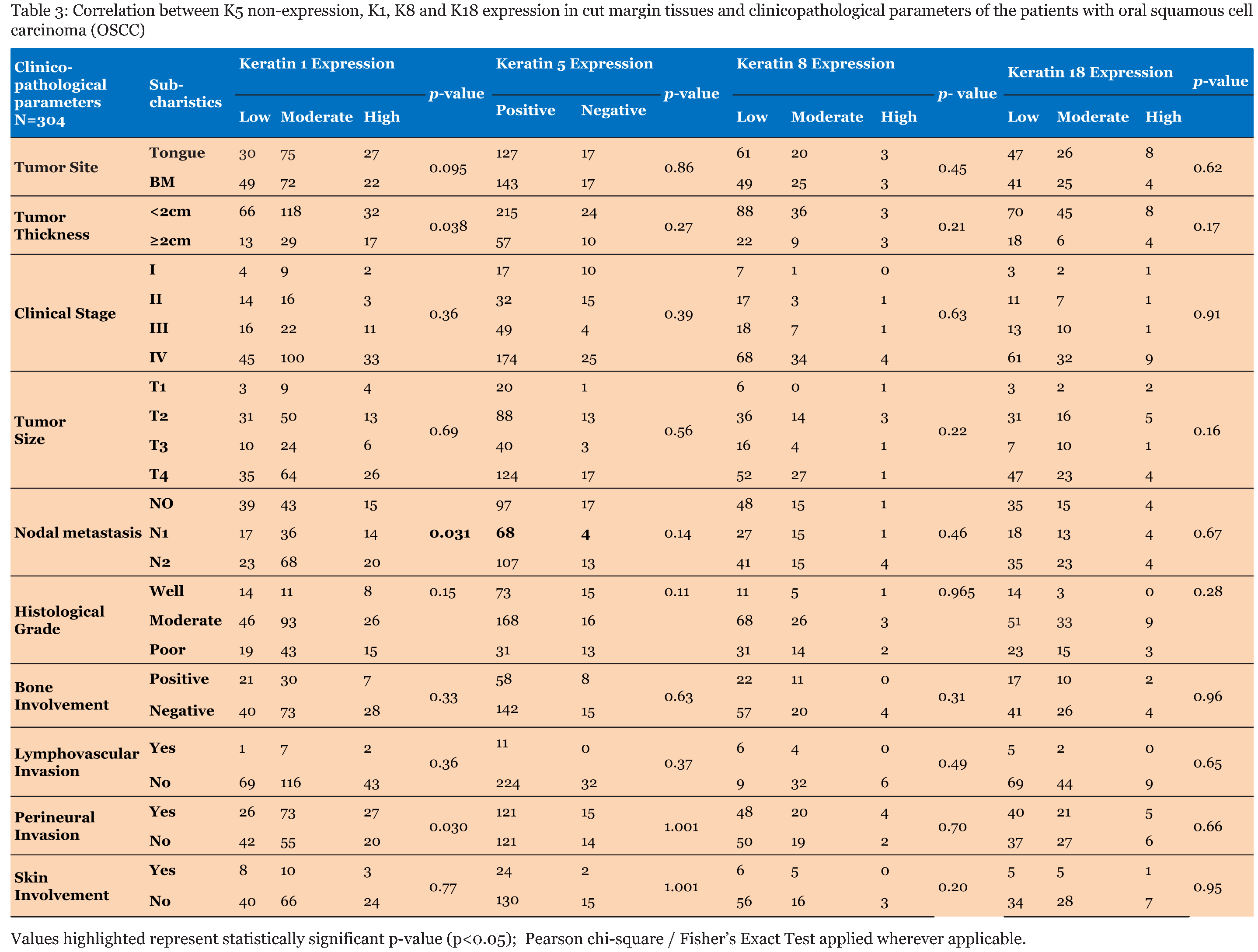
K5 expression was not detected in 8% of tongue and 11% of BM tumor tissues (Figure 2C). Their respective cut margin tissues also showed non-expression of K5 in 12% of tongue and 11% of BM tissues, respectively (Figure 2C). Different patterns of K5 immunolocalization in comparison with normal mucosal tissues were seen in hyperplastic/dysplastic, fibrotic as well as SCC tissues studied. Those types are i) Weak overall staining intensity for K5. ii) Loss of K5 staining in the basal and immediate suprabasal cell layers. iii) Loss of K5 staining in all the epithelial cell layers (Figure 2A:a-h). The staining intensity of K5 positive samples varied from sample to sample and sometimes even in the same tissue section, different cells stained with different intensity (Figure 2A:e-f, Figure 2D). K5 non-expression in tumor tissues significantly correlated with tumor site (p=0.023), nodal metastasis (p=0.029), and local recurrence (p=0.037), (Table 2) (Figure 2E). Although K5 expression in tumor tissues significantly correlated with nodal metastasis, its immunostaining was not detected in the lymphatic cells of either nodal metastatic or non-metastatic tumors (Figure 3:a, b). | ||||||
|
| ||||||
|
Kerating 1 expression | ||||||
|
| ||||||
|
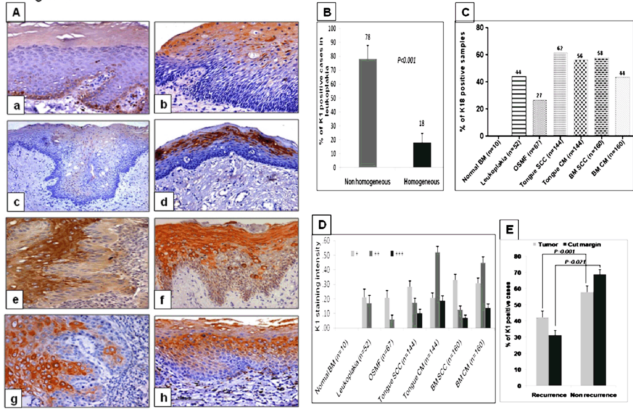
Detectable levels of K1 expression were also seen in 81/144 (56%) of tongue tumors and 143/160 (89%) of BM tumors while respective cut margin tissues of tongue 132/144 (92%) and BM 85/160 (53%) showed K1 immunolabeling (Figure 4C). Moderate intensity of K1 staining was seen in majority of samples while few samples also showed weak to intense staining (Figure 4D). K1 expression in tumor tissues significantly correlated with tumor size (p=0.030), nodal metastasis (p=0.040), bone involvement (p=0.007), skin involvement (p=0.040), and development of local recurrence (p=0.001) (Table 2, Figure 4E), while in cut margin tissues it correlated with tumor site (p=0.001), nodal metastasis (p=0.002), and perineural invasion (p=0.022). (Table 3) (Figure 4E). Immunolabeling for K1 was not detected in the lymphatic cells of nodal metastatic or non-metastatic tumors (Figure 3:c, d). | ||||||
| ||||||
|
Keratin 8 expression | ||||||
|
| ||||||
|
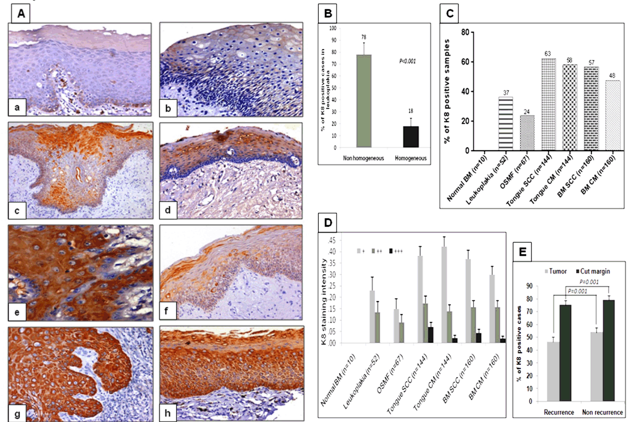
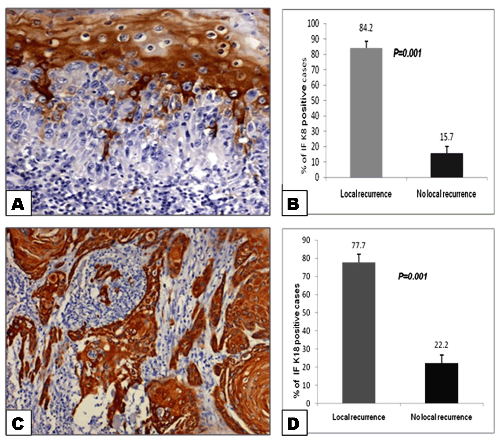
Detectable level of K8 expression was also seen in 90/144 (63%) of tongue and 91/160 (59%) of BM tumors while their respective cut margin tissues showed K8 labeling in 84/144 (57%) of tongue tissues and 76/160 (48%) of BM tissues (Figure 5C). Overall staining intensity for K8 was weak to moderate although few samples also showed intense labeling (Figure 5D). K8 expression in tumor tissues significantly correlated with tumor size (p=0.042), lymphovascular invasion (p=0.023) and local recurrence (p=0.001) (Table 2) (Figure 5). Its expression in cut margin tissues also significantly correlated with lymphovascular invasion (p=0.024) and local recurrence (p=0.001) (Table 3) (Figure 5E). It is a well-known fact that many times localization of protein can determine its function and thus may be of clinical importance. Hence, we analyzed K8 immunolabeling in the invasive front of tumor cells. K8 immmunolabeling was seen in the cells at tumor fronts of 70/181 (37%) invasive tumors (Figure 6A). Out of those 70 patients whose tumor invasive fronts were positive, 59 (84%) patients eventually developed recurrence (Figure 6B). Although, K8 immunolabeling was positive in tumor cells of nodal metastatic/non-metastatic SCC, it was not detected in the lymph nodes of same tumors (Figure 3:e,f). | ||||||
|
| ||||||
|
Keratin 18 expression | ||||||
|
| ||||||
|
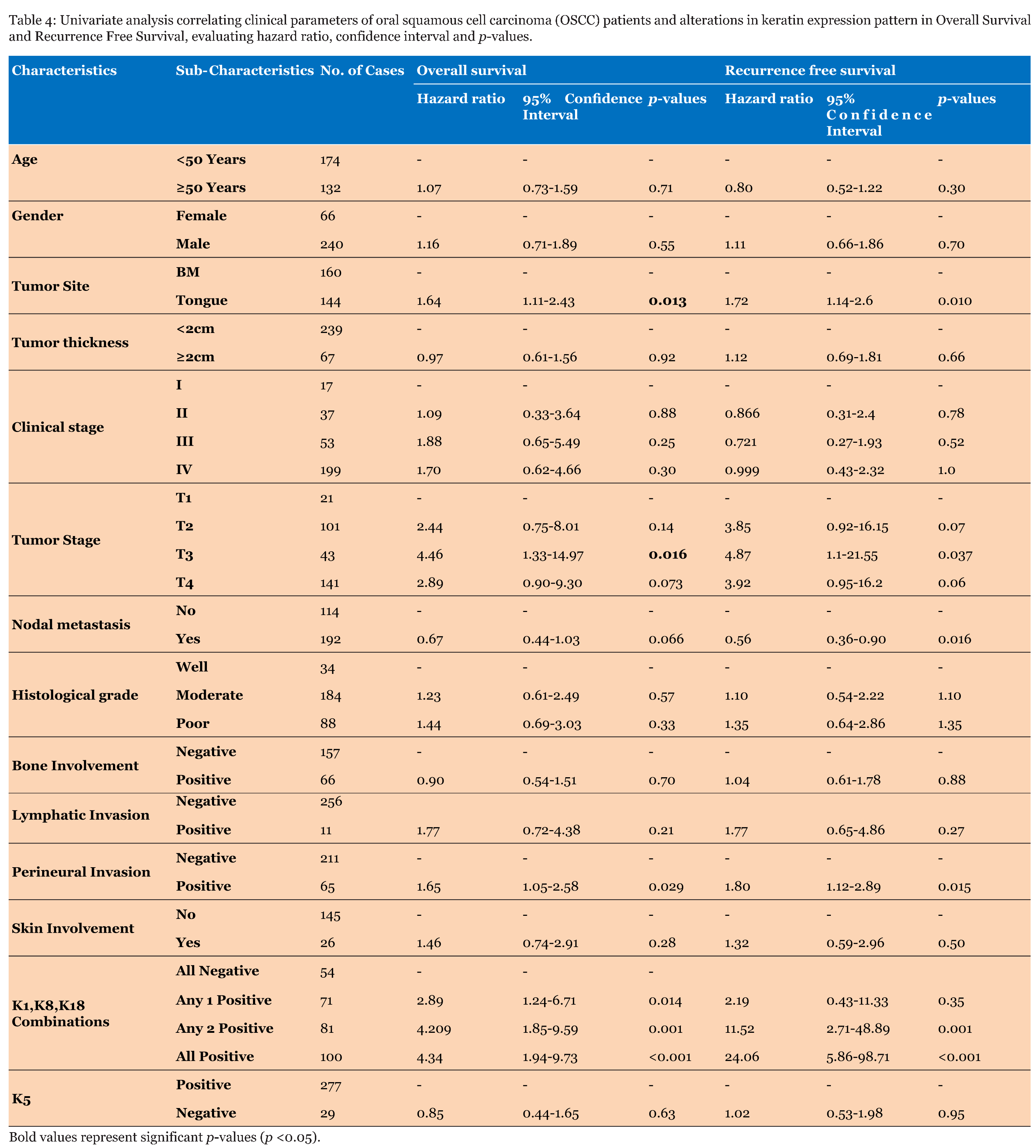
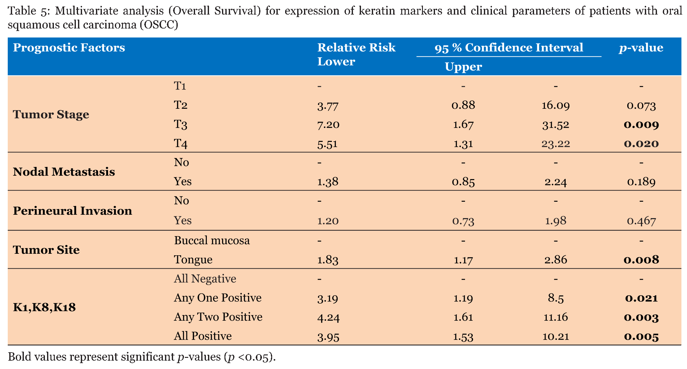
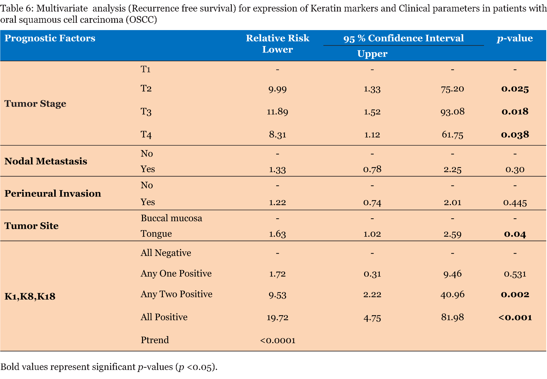
K18 immunostaining was found in 89/144 (62%) of tongue tumors and 93/160 (58%) of BM tumors while, their respective cut margin tissues showed K18 labeling in 81/144 (56%) of tongue tissues and 70/160 (44%) of BM tissues (Figure 7C). The overall staining intensity of K18 was moderate although few samples also demonstrated weak or intense labeling (Figure 7D). It is known that K8 and K18 are pairing partners, but in some of the tissues the immunolabeling for both K8 and K18 in the serial sections of the same tissue was not observed (Figure 5A: a, b, d, f, h) and (Figure 7A: a, b, d, f, h). K18 expression in tumor tissues significantly correlated with tumor stage (p=0.028), nodal metastasis (p=0.013), and local recurrence (p=0.001) (Table 2) (Figure 7E) while, in cut margin tissues its expression significantly correlated with development of local recurrence (p=0.001) (Figure 7E). K18 immunolabeling was seen in the invasive front of 81 tumor samples (Figure 6C). Out of these, 63 (78%) patients eventually developed recurrent tumor (Figure 6D). Although, K18 expression significantly correlated with nodal metastasis, it was not detected in the lymph nodes of the metastatic/non-metastatic tumor sections (Figure 3: g,h). Correlation between altered keratin expression pattern and survival of oral cancer patients: univariate and multivariate analysis | ||||||
|
| ||||||
| ||||||
| ||||||
|
| ||||||
|
Discussion
| ||||||
|
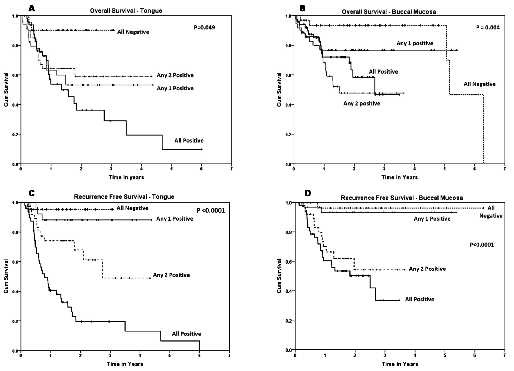
Keratins are epithelia-predominant intermediate filament proteins, which are expressed in a tissue-specific and paired manner [23]. However, alterations in their normal expression pattern have been shown in different pathological disorders, such as, gingivitis, psoriasis, and hyperkeratosis [24] [25]. Alterations have also been shown in leukoplakia, oral submucous fibrois (OSMF), and OSCC [26] [27]. In this study, immunohistochemistry was carried out to localize and semi-quantitate the expression of K1, K5, K8 and K18 in normal oral mucosa, oral epithelial hyperplasia/dysplasia, OSMF and OSCC tissues. The results of the IHC analysis were correlated with clinicopathological parameters of respective patients. In this study, we have made an attempt to cover major histopathological stages occurring during the process of oral tumor development. This study also covers two major sub-sites of the oral cavity that is tongue and BM which are the most prevalent sub-sites for the development of tumor in Indian patients due to the typical habits of chewing tobacco and allied products. These two sub-sites not only vary in their anatomical location and function but also show different pattern of keratin expression. The percentage of positivity and staining intensity of keratin expression was high for tongue tissues as compared to buccal mucosa. Overall survival rate for three years (52.3%) and five years (22.9%) in patients with tongue cancer was poorer as compared to patients with buccal mucosal cancer (66.8% and 66.8%, respectively). Also, it is general clinical observation that tongue tumors are more aggressive than the buccal mucosal tumors. Hence, we evaluated the significance of aberrantly expressed K1, K8 and K18 in different combinations with overall survival and recurrence free survival separately in subset of patients with tongue and buccal mucosal cancer. Interestingly, combinations of different keratin markers expressed by these sub-sites significantly correlated with overall survival as well as recurrence free survival. The expression of keratin pair 5/14 is a hallmark of squamous epithelium and is predominantly seen in the basal layers of stratified epithelium. This cell layer is mainly composed of tissue specific stem/progenitor cells. Stem cells of stratified epithelium have been described as the major cellular targets for cancer causing mutations and therefore might give in a long-term rise to the development of cancer. In this context, although K5 is a normal expression in oral cavity, it is interesting to evaluate the clinical significance of alterations in its expression pattern. In this study, we have observed different patterns of K5 immunolocalization in both premalignant SCC tissues as shown in results (Figure 2A: a-h). Previously, we have shown loss of K5 at protein [18] [19] [20] as well as RNA level [14] in oral premalignant lesions including in OSCC. We have also reported the role of K5/14 in regulation of cell differentiation/proliferation [28]. This keratin pair is known to negatively regulate cell differentiation. Thus it is possible that loss of K5 may induce changes which lead to abnormal cell differentiation. Other than us, only Morgen and Su [29] have shown loss of K5 expression in human oral dysplasia and SCC. Although, they have shown down regulation of K5 and K14 in some poorly differentiated SCC of oral cavity tissues, we have seen loss of K5 in all grades of SCC including hyperplasia and dysplasia. In this study, the immunoreactivity of all primary antibodies was tested on relevant frozen tissues to rule out the possibility of detection of artifacts due to formalin fixation. Further, we have correlated the non-expression of K5 with clinicopathological parameters of patients with precancerous lesions as well as SCC. K5 non- expression was seen in significantly higher number of non-homogenous leukoplakia samples as compared to homogeneous leukoplakia samples. Non-expression of K5 also significantly correlated with progressive grade of dysplasia. It is known that in Indian patients, the typical habits of chewing tobacco/areca nut and other ingredients contribute to the pathogenesis of leukoplakia and SMF [17] [30] [31]. Majority of the subjects recruited in this study had habits of chewing tobacco/areca nut along with other multiple habits. Thus K5 non-expression appears to be an early change occurring in the process of tobacco related oral carcinogenesis. Keratin pair of 1/10 is known to be a marker of cellular differentiation, and many well-differentiated SCCs derived from non-keratinizing stratified epithelia also express this keratin pair [18] [26]. In this study, cytoplasmic K1 expression has been detected in the intermediary cell layers of hyperplastic/dysplastic, fibrotic tissues as well as tumor tissues. The intermediary cells are usually comprised differentiated cell population. None of the sample showed K1 staining in the basal layer which is usually a proliferating cell population. K1 expression significantly correlated with some of the clinicopathological parameters of patients. For example, K1 aberrant expression was seen in significantly higher number of non-homogenous buccal leukoplakia samples as compared to homogeneous leukoplakia samples. It has been reported that non-homogenous leukoplakia is more prone for malignant conversion over a period of time as compared to homogenous leukoplakia [32]. A recent report on large-scale follow-up study also showed high risk of malignant transformation in patients who had non-homogenous leukoplakic lesions with high-grade dysplasia located at their lateral/ventral tongue [33]. K1 expression also significantly correlated with the progressive grade of dysplasia. The staining intensity as well as percentage of K1 positive SMF tissues increased with increase in the degree of fibrosis. These results collectively suggest that K1 expression levels are indicative of degree of dysplasia and fibrosis. Further, in oral SCC K1 expression significantly correlated with tumor size, nodal metastasis, bone and skin involvement and local recurrence and inversely correlated with patient's survival. However, Fillies group [15] did not find significant correlation between K1 expression and tumor size, nodal metastasis and patient's survival. Previously, K1 expression has been shown in well differentiated tumors and as the grade of the tumor increased, its expression was down regulated [26]. However, in this study we have detected K1 expression in the well, moderate as well as poorly differentiated tumors and intense immunolabeling was seen in higher grades of tumors. Thus, our results are at variance with reports from other laboratories. These differences probably can be explained by the fact that these reports are from the countries where tobacco chewing is not a major risk factor. Other important observation from our earlier studies was aberrant expression of K8 and/or K18 in precancerous lesions as well as SCC of oral mucosa [18] [19][20]. Glandular epithelia specific keratins 8/18 are normally not expressed in stratified epithelia [34]. We have shown the expression of this keratin pair in fetal buccal mucosa and tongue epithelium till 27 weeks of gestation [35]. As shown in case of K1, the aberrant expression of K8/18 also significantly correlated with non-homogeneous versus homogeneous leukoplakia. Further K8/18 expression also correlated with progressive grade of dysplasia and K18 with progressive grade of fibrosis. This indicates that aberrant expression of K8 and /or K18 could be used to assess the degree of dysplasia/fibrosis and may further be correlated with malignant potential of the tissues. It is known that SMF is a disorder of underlined connective tissue and it is proved that exposure of tobacco/areca nut causes epithelial cells to stimulate cytokines, and these cytokines are the real initiator of fibrosis [36]. Fibroblasts have been shown to be responsible for the structural and functional alterations of oral mucosa [37]. These reports suggest that both the compartments-connective tissue and epithelium have mutual influence on their cellular and functional regulators. Hence, occurrence of alterations in keratin expression pattern in fibrotic mucosa could be the cause of early molecular changes occurring in both the compartments. Our in vitro study has shown that K8/18 in some way contributes to the malignant transformation of stratified epithelial cells [38]. Results of the studies conducted by Casanova et al. [39] using K8 transgenic mice also support this finding. They have shown down-regulation of K5 in the epidermal cells where K8 transgene was expressed. Hence, it was important to analyze correlations between loss of K5 expression and gain of K1, K8, 18 expression with clinicopathological parameters of the patients to evaluate diagnostic and prognostic implications of these two events. However, in the present study K5 non- expression was seen only in 29/304 samples. Therefore, we have not compared K5 non-expression with aberrant expression of K1, K8 and K18. We have evaluated clinical significance of aberrantly expressed K1, K8 and K18 in combinations of any one positive, any two positive and all positive which significantly correlated with overall survival as well as recurrence free survival. We also noticed the trend that the risk of tumor recurrence increased, with increased number of markers combined together (Table 6). This indicates that a combination of these markers has better prognostic value as compared to any of these markers alone in OSCC patients. One of our important observations is presence of only one partner of this keratin pair in different subsets of cell populations of leukoplakia, SMF as well as SCC. Thus we see de-regulation of keratin pair expression in diseased condition. As is well known in keratin biology, keratins can form functional filaments only when they are expressed in specific pairs. Another observation in this study is the intense immunolabeling of K8/18 in the invasive front of the high grade tumors. It is known that several molecular events of importance for tumor spread occur at the tumor-host interface [40] . In this context, this observation is of particular importance to predict the biological aggressiveness of the tumor and could be taken into account during oral cancer management. As K5 non-expression and aberrant expression of K1, K8 and K18 were not detected in all the precancerous lesions, it was important to know whether those who demonstrated these two events are the high-risk lesions for malignant conversion over a period of time. However, majority of our patients with premalignant lesions were migrants from different states of India, with lack of stable contact details. Therefore, it was not possible for us to obtain the follow-up of the disease status of these patients. In this regard, it will be important to conduct further studies with systematic, periodic clinical follow-up of high risk oral cavity lesions. The altered keratin expression pattern was also seen in the cut margin tissues of the tumors, indicating that surrounding areas of the tumor although pathologically free from malignancy, show alterations at molecular level. In our previous study, we have found correlation between the expression of K8, K18 and K19 in the tumor surrounding areas and postsurgery elevated levels of their fragmented proteins in the sera of respective OSCC patients. This correlation showed association with development of local recurrence and poor survival of OSCC patients [21]. These observations further indicate that alterations in keratin expression pattern even in cut margin tissues may add value in histopathological diagnosis of progressive grade of OSCC. Thus in summary, we have detected altered K1, K5, K8 and K18 expression pattern in oral leukoplakia, submucous fibrosis and tumor tissues. Further, significant correlation between aberrant expression of K1, K8 and K18 and non-expression of K5 with clinical subtypes of leukoplakia was also seen. It is important to distinguish the homogenous and non-homogeneous leukoplakia lesions at early stage since it will help clinicians in determination of treatment protocols such as laser excision. We found statistical correlation between altered keratin expression patterns and histopathological progressive grade of dysplasia. At present histopathological diagnosis of dysplasia has remained subjective and such biomarkers will prove useful in minimizing this subjectivity especially when their presence or absence is important rather than their quantitation. Further, significant correlation was also found between the combinations of keratin markers-K1, K8 and K18 and overall survival as well as recurrence (local) free survival in patients with OSCC. Since tumor recurrence is one of the leading causes of death in oral cancer patients, this correlation will have important prognostic implications. | ||||||
|
Conclusion
| ||||||
|
This is the first comprehensive study evaluating statistical correlation between altered keratin expression pattern and clinicopathological parameters of patients with both oral potentially malignant lesions and malignant tumors. Our findings suggest that the alterations in keratin expression pattern may prove useful as surrogate markers for the diagnosis of oral potentially malignant disorders and may also have prognostic value in patients with oral cancer. | ||||||
|
Acknowledgements
| ||||||
|
We acknowledge grant support from Lady Tata Memorial Trust, Mumbai, India (Grant No. 1921). We thank Dr. Vaishali Natu from Nair Dental Hospital for providing leukoplakia and submucous fibrosis specimens and Dr. Swapna Deniz from D.Y.Patil Dental College and Hospital, Navi-Mumbai-India for providing leukoplakia specimens. We also thank Mrs. Namrata Mandavkar and Dr. Shilpi Sharma for retrieving patient's clinical information. | ||||||
|
References
| ||||||
| ||||||

|
[HTML Abstract]
[PDF Full Text]
|
|
Author Contributions:
Sharada Sawant – Conception and design, Acquisition of data, Analysis and interpretation of data, Drafting the article, Critical revision of the article, Final approval of the version to be published Milind Vaidya – Analysis and interpretation of data, Critical revision of the article, Final approval of the version to be published Devendra Chaukar – Analysis and interpretation of data, Critical revision of the article, Final approval of the version to be published Prakash Gangadaran – Acquisition of data, Critical revision of the article, Final approval of the version to be published Archana Kumari Singh – Acquisition of data, Critical revision of the article, Final approval of the version to be published Siddheshwar Rajadhyax – Acquisition of data, Drafting the article, Final approval of the version to be published Sadhana Kannan – Analysis and interpretation of data, Drafting the article, Final approval of the version to be published Padmavathi A. – Analysis and interpretation of data, Drafting the article, Final approval of the version to be published Shubhada Kane – Analysis and interpretation of data, Critical revision of the article, Final approval of the version to be published Sandeep Pagare – Analysis and interpretation of data, Critical revision of the article, Final approval of the version to be published Ranganathan Kannan – Analysis and interpretation of data, Critical revision of the article, Final approval of the version to be published Anil D'Cruz – Analysis and interpretation of data, Critical revision of the article, Final approval of the version to be published |
|
Guarantor of submission
The corresponding author is the guarantor of submission. |
|
Source of support
None |
|
Conflict of interest
Authors declare no conflict of interest. |
|
Copyright
© 2014 Sharada Sawant et al. This article is distributed under the terms of Creative Commons Attribution License which permits unrestricted use, distribution and reproduction in any medium provided the original author(s) and original publisher are properly credited. Please see the copyright policy on the journal website for more information. |
|
|